
Vertebral compression fractures
Kyphoplasty: stopping a broken vertebra from getting worse
A minimally invasive, outpatient procedure that stabilizes a fractured vertebra and relieves the pain of a collapsed spine bone — usually the same day, usually with a single small incision.
The problem
What a vertebral compression fracture is
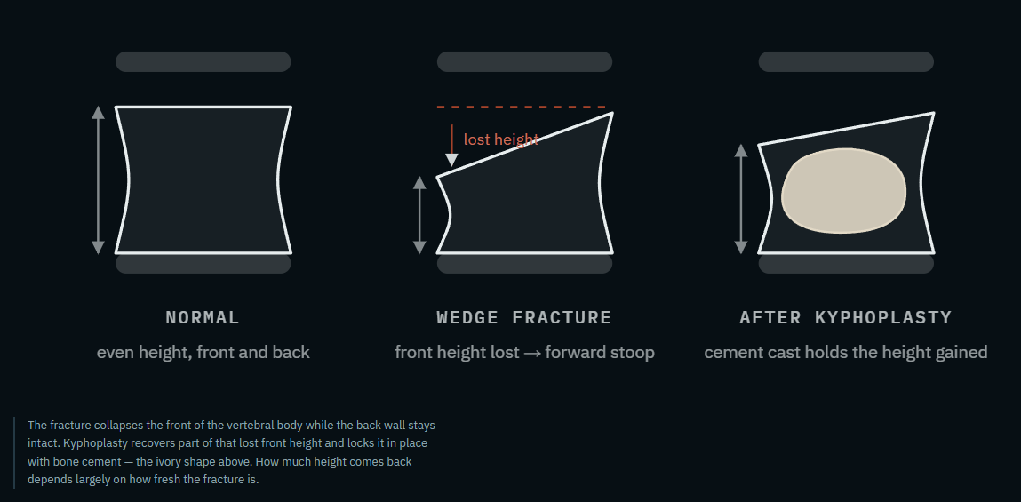
A vertebral compression fracture is a collapse of the front part of a spine bone. Most begin with osteoporosis: bone density has thinned to the point that ordinary daily loading — standing up, bending, a minor stumble, sometimes nothing identifiable at all — is enough to crush the front of the vertebra like an aluminum can. The vertebra does not shatter and displace; it compresses and wedges, which is why these fractures rarely injure the spinal cord directly, but they are still a structural failure of the bone.
Untreated, a compressed vertebra tends to keep settling under load. Each fracture increases the mechanical stress on the vertebrae immediately above and below it, which is part of why one compression fracture significantly raises the risk of another. Multiple fractures accumulate into the forward-stooped posture (kyphosis) sometimes called a dowager's hump, along with loss of height, and in more advanced cases, compression of the chest and abdomen that affects breathing and digestion.
The procedure
What kyphoplasty does
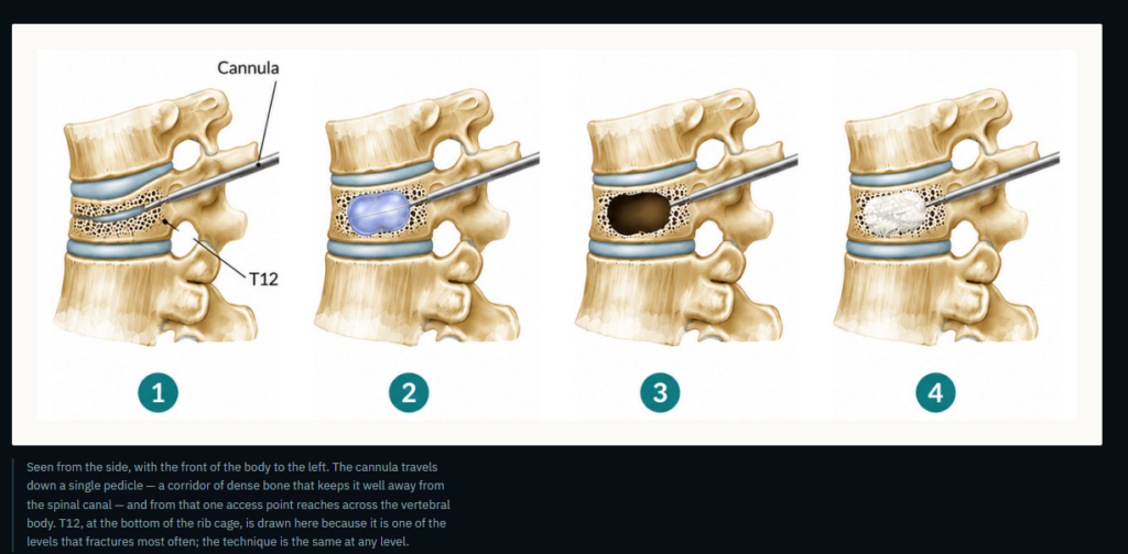
Kyphoplasty is done through the skin — there is no open incision, no muscle stripped off the spine, and no hardware left behind. Working under live X-ray guidance, a narrow needle is passed down the pedicle, the bony tunnel that connects the back of the vertebra to its body, and into the fractured bone. With the system currently used, one pedicle is enough: a single access point reaches across the vertebral body, so there is one small incision rather than two. A balloon is inflated inside the collapsed bone to lift the endplate and create a cavity. The balloon comes out, and that cavity is filled with acrylic bone cement, which hardens in minutes into an internal cast.
That cavity is the difference between kyphoplasty and plain vertebroplasty. Making a space first means the cement can be placed as a thick paste at low pressure, rather than injected as a thin liquid into porous bone — which is why cement leaks outside the vertebra are less frequent with kyphoplasty.
01
Access
One small skin nick. Under continuous fluoroscopy, a needle is advanced through the pedicle into the fractured vertebral body. Nothing crosses the spinal canal.
02
Inflate
A balloon tamp is expanded inside the collapsed bone, pushing the depressed endplate back up and compacting the surrounding bone into a firm-walled cavity.
03
Cavity
The balloon is deflated and removed, leaving a defined space. This is what lets the cement go in thick and slow rather than thin and pressurized.
04
Cement
Bone cement is delivered in small increments, watched on live X-ray the entire time. It hardens within minutes. The needle comes out; the skin usually needs no stitches.
Indications
Who kyphoplasty helps
Kyphoplasty works when it is aimed at the right fracture. The single most important question is not whether a fracture is visible on imaging — old, healed fractures are visible for life — but whether this fracture is the one generating this pain, right now.
- A painful vertebral compression fracture that is still biologically active, confirmed by marrow edema on MRI (STIR sequence) or uptake on a bone scan when MRI cannot be done.
- Pain that matches the level. Tenderness on examination at the fractured segment, not diffusely across the back.
- Pain severe enough to matter — limiting walking, sleep, or the ability to care for oneself, typically 5 or higher out of 10.
- A recent fracture. Most often within the past several weeks to a few months. Earlier treatment generally restores more height.
- Inadequate relief from conservative care — rest, bracing, and analgesics — or pain so severe from the outset that weeks of waiting is not a reasonable plan, particularly in an older adult for whom bed rest itself carries risk.
- Fractures from cancer or myeloma, where the bone will not heal on its own and waiting has no upside.
On the evidence: early sham-controlled trials of vertebral augmentation enrolled patients with fractures of any age and any pain severity, and found little benefit. Later trials that selected for acute, severely painful fractures — VAPOUR, VERTOS IV, and the FREE trial of balloon kyphoplasty — found clear advantages in pain and function. The lesson of that literature is not that the procedure works or does not work; it is that patient selection is the procedure.
Contraindications
When it is the wrong operation
Part of doing a high volume of these is turning them down. Kyphoplasty is not appropriate when:
- The fracture is old and healed, or found incidentally without matching pain.
- It is a burst fracture with bone displaced backward into the spinal canal.
- There is spinal cord compression, myelopathy, or a progressive neurological deficit — these need a surgical decompression, not cement.
- There is active infection: spinal osteomyelitis, discitis, or bacteremia.
- Bleeding cannot be safely controlled, or there is a known allergy to the cement components.
- The back pain is coming from something else — spinal stenosis, facet arthritis, or a disc — which is more common than patients expect and is why the MRI matters.
Seek care immediately, not an appointment, if back pain comes with new leg weakness, numbness in the groin or inner thighs, or loss of bladder or bowel control. That combination suggests pressure on the spinal cord or nerve roots and is treated as an emergency.
Spinal tumors
Fractures caused by cancer
Not every collapsed vertebra is osteoporotic. Multiple myeloma and metastatic disease — most often from breast, lung, prostate, kidney, or thyroid primaries — destroy the vertebral body from within, and those fractures do not heal with time and a brace. They tend to be relentlessly painful.
For these patients kyphoplasty is frequently combined with radiofrequency ablation. A small probe is placed into the tumor through the same access, and controlled heat is used to destroy tumor tissue within the bone; cement is then injected to restore structural support. The two work together: ablation addresses the tumor and the pain fibers, and the cement addresses the mechanical failure. Radiation therapy takes weeks to relieve pain and does nothing for the bone's strength, so ablation and augmentation are often used alongside it rather than instead of it.
This work is coordinated with medical and radiation oncology, and it can usually be done between systemic therapy cycles without interrupting cancer treatment.
What to expect
The day itself, and the days after
| Before | Nothing to eat after midnight. Blood thinners may need to be held — you'll be told exactly which and for how long. Bring your imaging if it was done elsewhere. |
| Arrival | You check in as an outpatient. An IV is placed, and you will meet the nurse and Dr. Fortes. |
| Sedation | Outpatient cases are done under moderate sedation with local anesthetic — comfortable and drowsy, breathing on your own, no breathing tube. |
| Procedure | Roughly 30 to 45 minutes for a single level, lying face down. You will hear the X-ray equipment moving; you will not feel the needle. |
| Recovery room | One to two hours flat, then sitting, then standing and walking with the nursing staff before discharge. |
| Same day | Every patient treated as an outpatient in our office goes home the same day. You will need a driver. The cement is already at full strength before you leave. |
| 24–48 hours | This is when most people notice the change — the sharp, movement-triggered pain is usually the first thing to go. Some soreness at the needle sites is normal and settles within a few days. |
| First week | Walking is encouraged from day one. Avoid heavy lifting and repeated bending. No wound care beyond keeping the sites dry for 24 hours. |
| Follow-up | A visit within two to four weeks to confirm the pain pattern has resolved, and to make sure the osteoporosis work-up is under way. |
Risks, stated plainly. The most common finding is cement extending slightly beyond the vertebral body, which is usually seen on imaging and causes no symptoms. Less common are infection, bleeding, nerve irritation, and a new fracture at an adjacent level. Rare but serious complications include cement entering a vein and travelling to the lungs, and injury to nerve tissue. Vertebral augmentation is a well-established procedure with a low complication rate in experienced hands, but it is not a zero-risk one.
Just as important
Kyphoplasty treats the fracture. It does not treat the osteoporosis.
A vertebral compression fracture is often the first hard evidence that bone density has been failing for years, and it is the strongest single predictor of the next fracture. Cementing one vertebra does nothing to protect the others.
So every osteoporotic fracture patient should also leave with a bone-health plan: a DEXA scan, vitamin D and calcium checked and corrected, a review of medications that weaken bone, a fall-safety look at the home, and — for most people after a fragility fracture — prescription therapy to rebuild or preserve bone density. That part happens with your primary physician or an endocrinologist, and the referral is made rather than assumed.
Experience
Why experience matters here
Kyphoplasty looks simple on a diagram and is not simple in practice. The judgment sits in the parts a diagram cannot show: reading which level on the MRI is actually generating the pain when three vertebrae look abnormal; choosing a needle trajectory in a pedicle that osteoporosis has left narrow and soft; knowing when the cement is at the right viscosity to advance and when to stop; and recognizing the case that should be sent to a spine surgeon instead.
Questions patients ask
Will I be asleep? Where is it done?
Not fully asleep. All outpatient kyphoplasties are performed under moderate sedation with local anesthetic — comfortable and drowsy, breathing on your own, with no breathing tube. They are done in our Orlando office at Orlando Neurosurgery, 265 E Rollins Street, Suite 12000, Orlando, FL 32804, and you go home the same day.
How long does the pain relief last?
The cement is permanent and does not wear out. When the treated fracture was the source of the pain, the relief is durable. What can happen later is a new fracture at a different level — which is a different problem, and the reason osteoporosis treatment matters as much as the procedure.
Is it too late? My fracture is a few months old.
Possibly not. What matters is whether the fracture is still active on MRI. Some fractures remain unhealed and painful for many months, and those still respond. If the MRI shows the bone has healed, cement will not help and you will be told so.
Will I be taller? Will the hump go away?
Some height is usually recovered, more so in fresher fractures, but kyphoplasty is a pain and stability procedure rather than a cosmetic one. Its real value in posture is preventing further collapse rather than reversing what has already happened.
Can more than one level be treated at the same time?
Yes. Two or three levels in one session is common when each is confirmed to be an active, painful fracture. Not every abnormal-looking vertebra needs treating — only the ones the imaging and the examination both point to.
What about the metal detector at the airport?
Bone cement is acrylic, not metal. It will not set off a detector, and it does not restrict future MRI scans.
Our office
Scheduling and referrals
Location
Orlando Neurosurgery
265 E Rollins Street, Suite 12000
Orlando, FL 32804
Contact
Phone: 407-975-0200
Fax: 407-975-0209